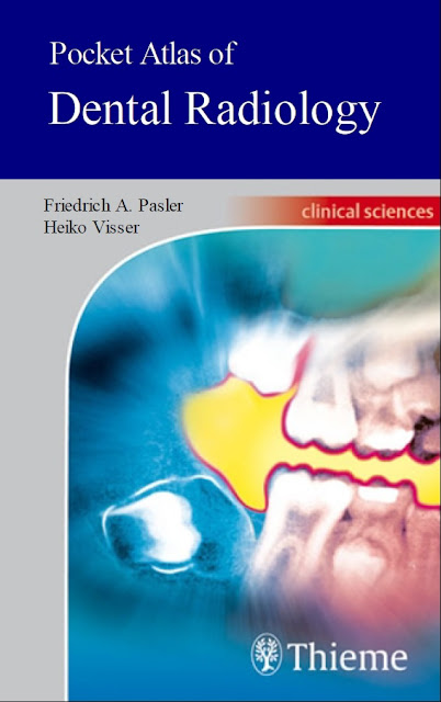
In this age of highly specialized medical imaging, an examination of the teeth and alveolar bone is almost unthinkable without the use of radiographs. This highly informative and easy-to-read book with a collection of 798 radiographs, tables, and photos provides a myriad of problem-solving tips concerning the fundamentals of radiographic techniques, quality assurance, image processing, radiographic anatomy, and radiographic diagnosis. Information is easy to find, enabling the reader to literally get a grasp of essential new knowledge in next to no time. The dental practice team now has a pocket consultant at its fingertips, providing practical ways to incorporate new techniques into daily practice. A fine-tuned didactic concept: Each topical concept is printed compactly on a double-page spread on the left: concise and highly instructive text on the right: informative, high-quality illustrations
Main topics include:
- Examination strategies, radiation protection, quality assurance
- Conventional and digital radiographic techniques
- Radiographic anatomy: The problems of object localization and how to solve them
- Recent research with conventional radiography, CT, MRI, etc.
- Normal variations and pathologic conditions as viewed with the various imaging techniques
- A concise and up-to-date presentation of modern dental radiology
Editorial Reviews
From the Back Cover
In this age of highly specialized medical imaging, an examination of the teeth and alveolar bone is almost unthinkable without the use of radiographs. This highly informative and easy-to-read book with a collection of 798 radiographs, tables, and photos provides a myriad of problem-solving tips concerning the fundamentals of radiographic techniques, quality assurance, image processing, radiographic anatomy, and radiographic diagnosis. Information is easy to find, enabling the reader to literally get a grasp of essential new knowledge in next to no time. The dental practice team now has a pocket consultant at its fingertips, providing practical ways to incorporate new techniques into daily practice.
A fine-tuned didactic concept: Each topical concept is printed compactly on a double-page spread on the left: concise and highly instructive text on the right: informative, high-quality illustrations
Main topics include:
- Examination strategies, radiation protection, quality assurance
- Conventional and digital radiographic techniques
- Radiographic anatomy: The problems of object localization and how to solve them
- Recent research with conventional radiography, CT, MRI, etc.
- Normal variations and pathologic conditions as viewed with the various imaging techniques
- A concise and up-to-date presentation of modern dental radiology
About the Author
Heiko Visser is Professor, Dental School, University of Gottingen, Gottingen, Germany.
Department of Periodontology, Institute of Dental and Oral Medicine, Goettingen, Germany
- Name of Book: Pocket Atlas of Dental Radiology
- Format: pdf
- Categories: Dentistry (General)
- Writer(s): Heiko Visser
- Publisher: Thieme
- File Size: 87.5mb
Oral Rehabilitation with Dental Implants Volume 3 PDF Free Download
Pocket Atlas of Dental Radiology PDF Free Download
Alright, now in this part of the article, you will be able to access the free download of Pocket Atlas of Dental Radiology using our direct links mentioned at the end of this article. We have uploaded a genuine PDF ebook copy of this book to our online file repository so that you can enjoy a blazing-fast and safe downloading experience.
