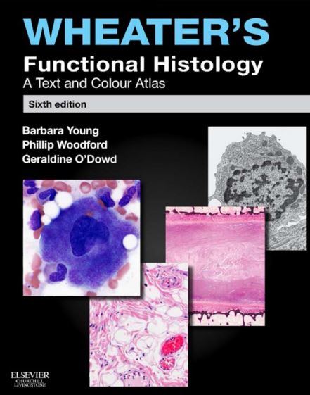
Wheater’s Functional Histology: A Text and Colour Atlas
In this article, we are sharing with our audience the genuine PDF download of Wheater’s Functional Histology: A Text and Colour Atlas PDF using direct links which can be found at the end of this blog post. To ensure user-safety and faster downloads, we have uploaded this .pdf file to our online cloud repository so that you can enjoy a hassle-free downloading experience.
At USMLE Med, we believe in quality and speed which are a part of our core philosophy and promise to our readers. We hope that you people benefit from our blog! Now before that we share the free PDF download of Wheater’s Functional Histology: A Text and Colour Atlas PDF with you, let’s take a look into few of the important details regarding this ebook.
Histology is a very colorful subject because of its diverse coloring combinations stained by hematoxylin and eosin staining media. Besides, this histology subject is very difficult to memorize because of its very slight detailing features. There are very insignificant details of histology that are mandatory to remember for studying purposes. It is very difficult to remember all important histological structures in any specific structure. This book, Wheater’s functional histology, ensures all the necessary details regarding the histology of different structures in a very unique and effective presentation. This book is known as the colorful atlas with text explanation for histology.
Medical students need to learn a bundle of books to learn the complete essential anatomy of the body structures. This book explains all the fundamental details of histology in a very straightforward manner. In addition, this book summarizes the important details of diagrams in the form of captions and keynotes. This book is a complete package of important highlights in the forms of text and further diagrammatic explanations for the sake of students.
These three worked hard, compiled all the text and images data, and organized all the details in a book form.
Barbara young
- Director of anatomical pathology, john hunter hospital
- Conjoint associate professor at University of Newcastle, new south wales, Australia
Geraldine O’Dowd
- Lead the author team for the recent edition of wheater’s functional histology
- Consultant diagnostic pathologist at Lanarkshire NHS Board
- An honorary clinical senior professor at the University of Glasgow, Glasgow
Phillip Woodford:
Senior staff specialist (anatomical pathology and cytopathology) at john hunter hospital, new castle, new south wales, Australia
Important target points in Wheater’s functional histology: A text and color atlas
This book, Wheater’s functional histology, highlights the main points of histology for the ease of medical students. These points are mentioned below:
Recognize histological details
This book enables the students to understand the histological features. In addition, it highlights the special diagnostic features of the cells present in any structure. This feature helps the students recognize these microscopic details in any clinical exam. Moreover, it also helps to provide all necessary information regarding international exams.
Clinical correlation
This book integrates the clinical details into the text to highlight the importance of histological features in clinical practice. In addition, this feature is helpful to establish a strong diagnostic assessment.
Staining techniques
At the end of the book, this book also explains the staining techniques and their result. This thing provides a strong basis for medical students to further establish powerful dimensions of histology.
Features of the 6th edition of Wheater’s functional histology: A text and color atlas
These are some of the salient features focused on in the 6th edition of Wheater’s functional histology. Moreover, these key aspects are helpful for medical students to recognize all the microscopic details during their clinical exams or laboratory practice. Some of these features are below:
- This book highlights all the important features necessary for medical students to recognize microscopic structures without any inconvenience.
- This book provides clinical correlation in the text to assess how to apply histology during any clinical or lab examination procedures.
- This book also provides rich details of images in the form of captions written below the images.
- This histology concise book is also available with the student consult. Moreover, this feature will provide complete access to all images, virtual histolab, self-assessment questions, and rationales.
- An eBook is available that includes multiple-choice questions for the assessment of medical students.
EBook features
This eBook feature includes:
- Highlighting the content, take notes, and search in the book
- May create digital flashcards instantly
- Use x-ray for important concepts ( x-ray provides instant access to all important terms and concepts in the book with glossary definitions, links to relevant textbook pages, related content from Wikipedia and youtube)
Content details of wheater’s functional histology: a text and color atlas
This book, Wheater’s functional histology, covers all important topics from the cell level to the organ level. At the start of the book, the authors compiled all the basic information and histology, physiology, and anatomy of cells. In this way, it starts from the minor level of organization and explains the basic histology concepts.
Moving forward, it highlights the important histologic features of all tissue forms. This book covers all the important histopathological features of the blood, bone marrow, connective, muscle, and nervous tissue. Moreover, this book not only explains the histology of these body tissues but also correlates the clinical aspects of histology with its histologic features.
After a thorough review of the tissue level, this book explains the major organ level organization that collaborates to form major important systems. This book furnishes all the histologic features of the important body systems. The major systems are:
- Skin
- Circulatory, skeletal, nervous, immune, respiratory, gastrointestinal, liver, urinary, and endocrine system
- Special emphasis on histology of the oral cavity, which is a major portion for dental students as well.
| Book name: | Wheater’s functional histology: A text and color atlas |
| Author: | Barbara Young Phillip Woodford Geraldine O’Dowd |
| Edition: | 6th |
Disclaimer:
This site complies with DMCA Digital Copyright Laws. Please bear in mind that we do not own copyrights to this book/software. We are not hosting any copyrighted contents on our servers, it’s a catalog of links that already found on the internet. Usmlemed.com doesn’t have any material hosted on the server of this page, only links to books that are taken from other sites on the web are published and these links are unrelated to the book server. Moreover Usmlemed.com server does not store any type of book, guide, software, or images. No illegal copies are made or any copyright © and / or copyright is damaged or infringed since all material is free on the internet. Check out our DMCA Policy. If you feel that we have violated your copyrights, then please contact us immediately.
![All MBBS PDF Books Free Download [First Year to Final Year] Download All MBBS Years Books](https://usmlemed.com/wp-content/uploads/2023/08/All-MBBS-PDF-Books-Free-Download-First-Year-to-Final-Year.jpg)
![Coloring Atlas of the Human Body PDF Free Download [Direct Link] Coloring Atlas of the Human Body PDF Free](https://usmlemed.com/wp-content/uploads/2023/09/Coloring-Atlas-of-the-Human-Body-PDF-Free-Download-Direct-Link.jpg)
![Gray’s Anatomy Coloring Book PDF Free Download [Direct Link] Gray’s Anatomy Coloring Book PDF Free Download](https://usmlemed.com/wp-content/uploads/2023/09/Grays-Anatomy-Coloring-Book-PDF-Free-Download-Direct-Link.jpg)
![Sobotta Atlas of Human Anatomy [All Volume] Sobotta Atlas of Human Anatomy [All Volume]](https://usmlemed.com/wp-content/uploads/2023/08/Sobotta-Atlas-of-Human-Anatomy-All-Volume.jpeg)
![Download USMLE Step 1 NBME Images 2023 PDF Free [Direct Link] Download USMLE Step 1 NBME Images 2023 PDF Free [Direct Link]](https://usmlemed.com/wp-content/uploads/2023/08/Download-USMLE-Step-1-NBME-Images-2023-PDF-Free-Direct-Link.png)
![Download (FREE) MedStudy Internal Medicine Videos 2023 Download (FREE) MedStudy Internal Medicine Videos 2023 [Direct Link]](https://usmlemed.com/wp-content/uploads/2023/09/Download-FREE-MedStudy-Internal-Medicine-Videos-2023.webp)
![First Aid for the USMLE Step 1 2023 33rd Edition PDF Free Download [Direct Link] First Aid for the USMLE Step 1 2023 33rd Edition](https://usmlemed.com/wp-content/uploads/2023/03/First-Aid-for-the-USMLE-Step-1-2023-33rd-Edition-PDF-Free-Download.png)